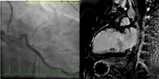
Fig 1.
A – coronary angiogram of the left coronary artery showing a proximal occlusion of the left anterior descending coronary artery with visible collaterals from the normal circumflex; B – magnetic resonance imaging reveals subendocardial late gadolinium enhancement in the apical and mid anterior and anteroseptal wall segments of the left ventricle with more than 50% of viable myocardium.

