
Fig 2.
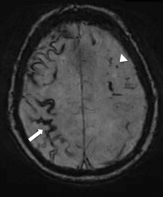
Cerebral amyloid angiopathy. Magnetic resonance susceptibility-weighted imaging sequence showing superficial siderosis (white arrow) and cerebral microhaemorrhage (white arrow head), which are features supportive of a diagnosis of cerebral amyloid angiopathy.