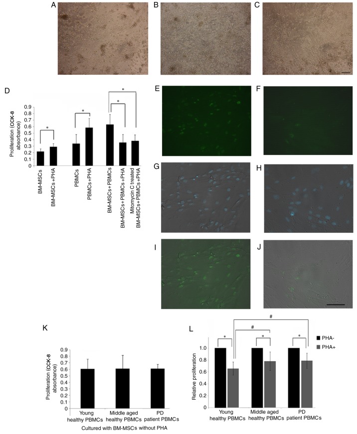
Figure 2.
Proliferation suppression of PBMCs by PHA-treated BM-MSCs. (A) PBMCs isolated from young healthy individuals. (B) Co-culture of BM-MSCs and PBMCs without PHA at day 3. (C) Co-culture of PHA-activated PBMCs and BM-MSCs at day 3. (D) In the presence of PHA, the proliferation of PBMCs and BM-MSCs increased significantly (*P<0.05). When incubated with BM-MSCs and mitomycin C-treated BM-MSCs, PHA-activated PBMC proliferation decreased significantly. Ki67 staining of the MSCs prior to (E, G and I) and following mitomycin C treatment (F, H and J) were demonstrated. (E and F) Ki67 staining. (G and H) Merged image of DAPI nucleus staining and phase contrast. (I and J) Merged image of Ki67 and phase contrast. (K) Without PHA, the PBMC proliferation in the young healthy, the middle-aged healthy, and the middle-aged PD individuals was not significantly different. (L) Following treatment with PHA, the PBMC proliferation in the young healthy, the middle-aged healthy and the middle-aged PD groups decreased significantly (*P<0.05). The proliferation of PBMCs suppressed by BM-MSCs of the young healthy subjects was significantly lower than that of the middle-aged healthy and the PD individuals (#P<0.05). Scale bar, 50 µm, n=6. PBMCs, peripheral blood mononuclear cells; PHA, phytohemagglutinin; BM-MSCs, bone marrow-derived mesenchymal stem cells; PD, Parkinson's disease.

