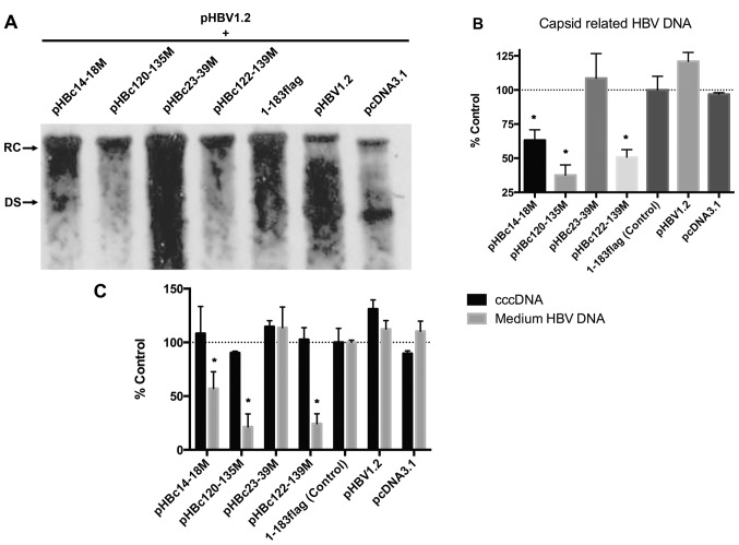
Figure 4.
HBc dimer-dimer mutants inhibit WT replication. (A) HepG2 cells were co-transfected with an equal amount of HBc mutant and pHBV1.2. After 5 days post-transfection, the capsid-associated HBV DNA was isolated and detected with a specific HBV DNA probe. (B) Relative density of the RC and DS bands of each group in (A) was analyzed using Quantity One software (version 4.6.3). The experiment was repeated in triplicate. Date are presented as the mean ± standard error of the mean. *P<0.05 vs. 1–183 flag control group. (C) After 5 days post co-transfection, the cell medium was collected and then purified with a 0.45-µm filter; the nuclear cccDNA was also purified. Nuclear cccDNA and virion HBV DNA in the culture medium were quantified by qPCR on a LightCycler® 480 system. The experiment was repeated in triplicate. Date are presented as the mean ± standard error of the mean. *P<0.05 vs. 1–183 flag control group. DS, double-stranded DNA; HBV, hepatitis B virus; HBc, HBV core protein; RC, relaxed circular DNA.

