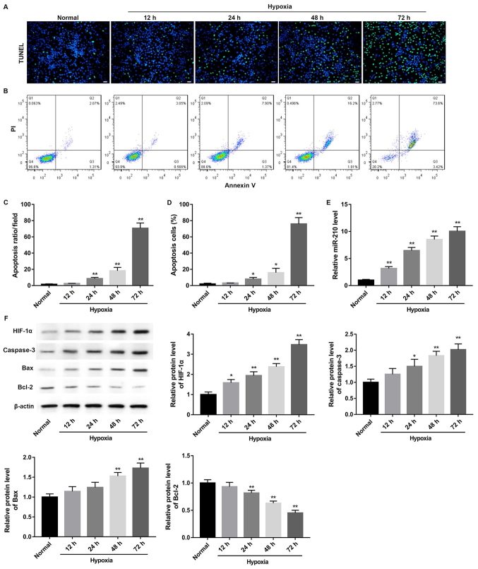
Figure 1.
Hypoxia induces apoptosis of GC-2 cells at different time points. (A) TUNEL staining results of GC-2 cells. Scale bar, 20 µm. (B) Representative graphs of flow cytometry analysis. (C) Apoptosis rate of GC-2 cells was evaluated by TUNEL staining. (D) Apoptotic GC-2 cells were measured by flow cytometry. (E) Reverse transcription-quantitative polymerase chain reaction analysis of miR-210 expression in mouse GC-2 cells subjected to hypoxia for 12, 24, 48 and 72 h. (F) Western blot analysis for HIF-1α, caspase-3, Bax and Bcl-2 protein expression in mouse GC-2 cells subjected to hypoxia for 12, 24, 48 and 72 h. *P<0.05, **P<0.01 vs. respective normal. miR, microRNA; HIF-1α, hypoxia-inducible factor-1α; Bax, apoptosis regulator BAX; Bcl-2, B-cell lymphoma 2; TUNEL, terminal deoxynucleotidyl-transferase-meditated dUTP nick end labeling and flow cytometry; PI, propidium iodide; GC-2, GC-2spd.

