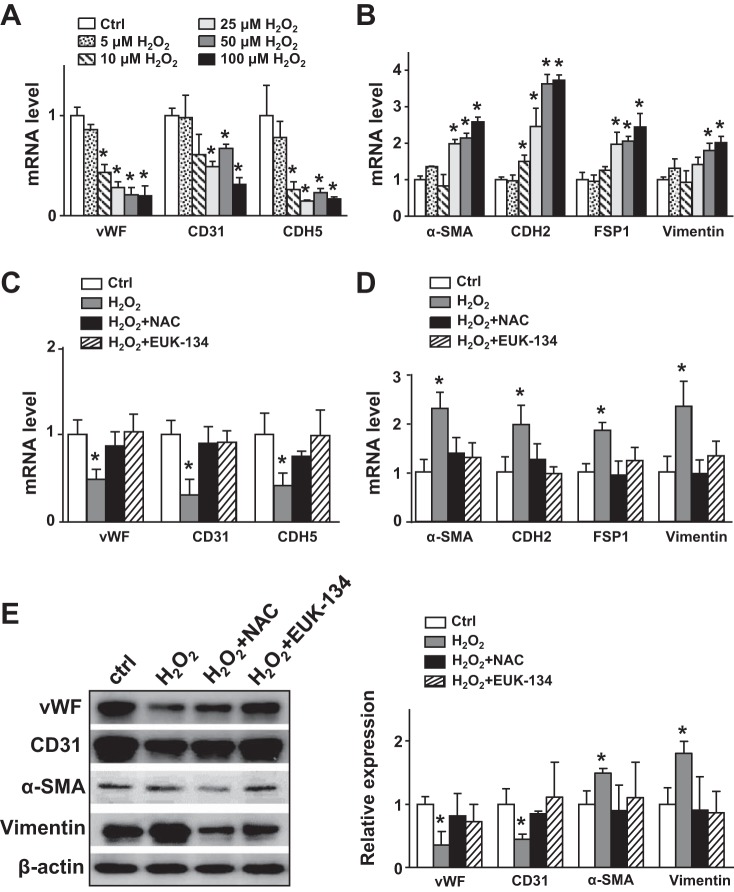
Fig. 3.
Oxidative stress in endothelial cells (ECs) promotes the endothelial-to-mesenchymal transition. A and B: human umbilical vein ECs were treated with H2O2 (5, 10, 25, 50, and 100 μmol/l) for 24 h. C–E: human umbilical vein ECs were pretreated with N-acetylcysteine (NAC; 5 mmol/l) or EUK-134 (1 μmol/l) for 5 h before incubation with H2O2 (100 μmol/l) for 18 h. mRNA levels of von Willebrand factor (vWF), CD31, cadherin 5 (CDH5, α-smooth muscle actin (α-SMA), cadherin 2 (CDH2), fibroblast-specific protein 1 (FSP1), and vimentin were measured by real-time quantitative PCR. E: protein levels of vWF, CD31, α-SMA, and vimentin were measured by Western blot analysis. Ctrl, control. Data are means ± SE from at least three independent experiments. *P < 0.05 between the indicated groups.

