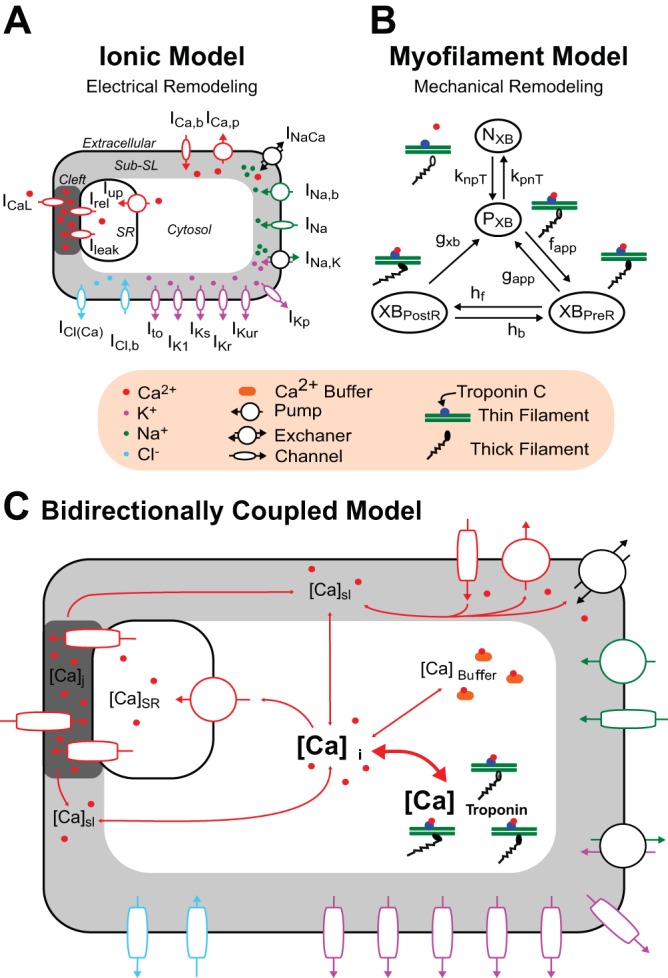
Fig. 1.

Bidirectionally coupled electromechanical model of the atrial myocyte. The human atrial ionic model by Chang et al. (5) (A) and the human myofilament model by Zile and Trayanova (44, 45) modified to match human atrial force data (B) were bidirectionally coupled (C) by having thin filament activation depend on free intracellular Ca2+ concentration ([Ca2+]i) and by incorporating mechanoelectric feedback via [Ca2+]i buffering of troponin C [thick double-headed red arrow linking [Ca2+]i and total Ca2+ bound to troponin C ([Ca2+]troponin)]. NXB and PXB are thin filament states where cross-bridge (XB) formation is inhibited (NXB) and where weakly bound XB formation is possible (PXB).
