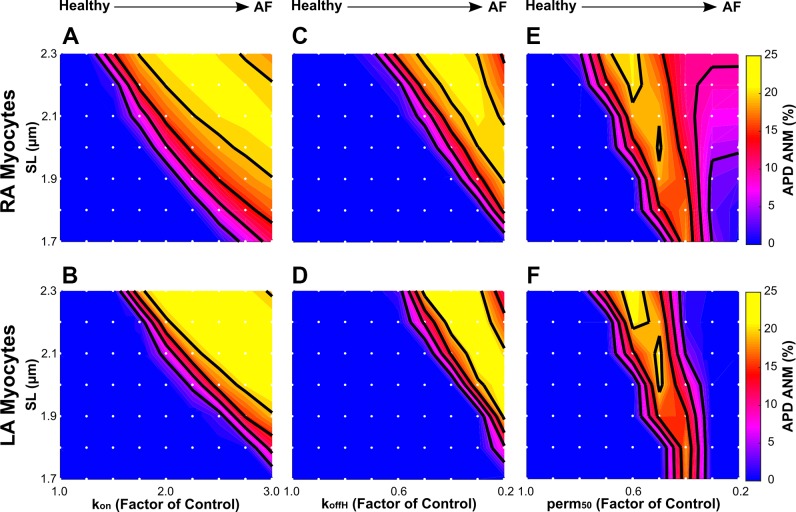
Fig. 7.
Sensitivity of action potential duration alternans-normalized magnitude (APD-ANM) to sarcomere length (SL) in simulations with our right atrial (RA; A, C, and E) and left atrial (LA; B, D, and F) electromechanical myocyte models with chronic atrial fibrillation (cAF)-induced ionic remodeling and varying degrees of cAF-induced myofilament remodeling. cAF-induced myofilament remodeling was incorporated as in Fig. 5. Each white dot represents a unique simulation in which the model was isometrically paced at a specific SL ranging from 1.7 to 2.3 µm in increments of 0.1 µm. Colors represent APD-ANM. Contour lines were drawn at APD-ANM of 5%, 10%, 15%, and 20%.

