
Figure 1.
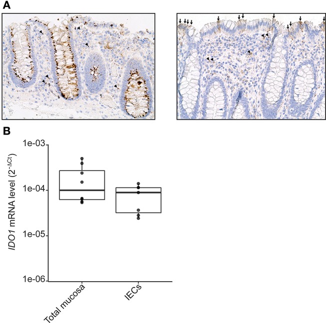
IDO-1 expression in human colonic epithelial cells. (A) Human normal colonic mucosa was stained for IDO-1. Representative immunohistochemical staining of IDO-1 showed that IDO-1 (brown) is expressed in epithelial cells [left: strong perinuclear and/or membrane staining of about 80% of the IECs; right panel: heterogeneous staining of few IECs (arrows)] and in few lamina propria mononuclear cells (arrowheads) and endothelial cells (asterisk) (original magnification × 200). (B). IDO-1 gene expression was determined by RT-PCR on RNA extracted from preparations of isolated human colonic epithelial cells (IECs) and of whole mucosa microdissected from normal colon. Results were normalized to β-2 microglobulin (B2M) and expressed as 2-ΔCt relative value (median ± quartiles) of 4 patients (1–2 samples/patient).