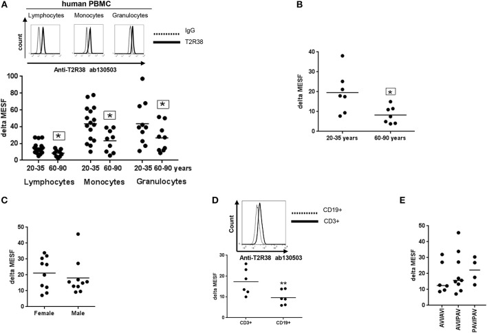
Figure 1.
Expression of T2R38 receptor in human PBMC. (A) Age-related T2R38 expression in PBMC from young (20–35 years, n ≥ 10) and elderly (60–90 years, n = 9) subjects. A representative staining from one person is shown for each cell type; T2R38 (continuous line) and rabbit IgG (dotted line). (B) T2R38 expression in CD3+ T cells from young (n = 7) and elderly (n = 7) donors (C) Sex difference of T2R38 expression in lymphocytes from male and female donors (n = 10). (D) T2R38 expression in CD3-PE stained T cells and CD19-APC stained B cells from human volunteers, (n = 6). A representative staining from one person is shown; CD3+ (continuous line) and CD19+ (dotted line). (E) Haplotype analysis was done using amplification of a TAS2R38 864bp fragment followed by sequencing. PAV: proline, alanine and valine; AVI: alanine, valine and isoleucine (n = 19). T2R38 expression (delta MESF) was quantified by Quantum Alexa Fluor 488 MESF using cytofluorometry (Supplementary Figure S1A), IgG was used as isotype control. Each dot presents one donor in the graphs. The gating strategy is shown in Supplementary Figure S1B. Significance of difference was calculated relatively to the respective control, *p < 0.05, **p < 0.01.

