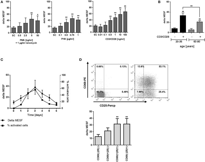
Figure 2.
Effect of activation on T2R38 expression in PBMC. (A) PBMC were stimulated with different concentrations of PMA/ionomycin (1 μg/ml), PHA or anti-CD3/CD28 mAbs for 72 h (n ≥ 5) (B) Isolated PBMC from young and elderly individuals were stimulated with 1 ng/ml CD3/CD28 for 72 h (n ≥ 5) (C) PBMC were stimulated with 1 ng/ml CD3/CD28 for the indicated time points, activated cells were determined by the percentage of blast cells (n ≥ 3) (D) PBMC were stimulated with 1 ng/ml CD3/CD28 and T2R38 expression on CD69+/CD25+ CD3+T lymphocytes determined at the indicated time points (n = 5). A representative staining of surface markers CD69+/CD25+ at day 2 from one subject is shown as scattergram. T2R38 expression (delta MESF) was assessed by Quantum Alexa Fluor 488 MESF beads in comparison to rabbit IgG isotype control; bars are means + SD (A,B) or means ± SD (C,D). Significance of difference was calculated relatively to the respective control, *p < 0.05; **p < 0.01. SC, solvent control. The gating strategy is shown in Supplementary Figure S2.

