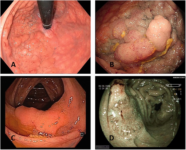
Figure 1.
(a) Endoscopic image of granular homogenous laterally spreading tumour (LST-G-H) with white light; (b) granular nodular mixed laterally spreading tumour (LST-G-N) with white light; (c) non-granular flat laterally spreading tumour (LST-NG-F) with white light; (d) image of non-granular pseudodepressed laterally spreading tumour (LST-NG-PD) with blue light imaging (BLI).

