
Fig. 1.
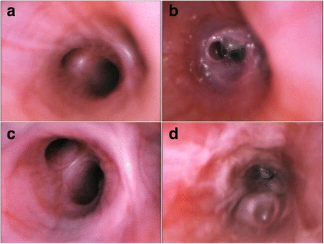
Bronchoscopic evaluation of mechanically ventilated Large White Landrace pigs challenged by Pseudomonas aeruginosa. a Main right upper bronchus, prior to bacterial challenge; of note no abnormalities can be found. b After 24 h from inoculation of 15 mL of 107 colony forming units of P. aeruginosa, the distal portion of the right middle bronchus is copiously filled with purulent secretions with a reduction of the distal bronchi by more than 60%. c Main right medium bronchus, prior to bacterial challenge, with no abnormalities. d After 24 h from inoculation of 15 mL of 107 colony forming units of P. aeruginosa, the bronchial mucosa is highly hyperemic and retained purulent secretions are evident throughout the bronchus, almost completely obstructing distal bronchi