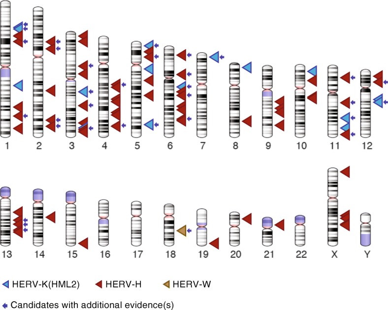
Fig. 5.

Karyotypic view of the location of the candidate dimorphic HERVs. The dimorphic candidates of HERV-K (HML2) are shown as blue triangles, HERV-H as red triangles and HERV-W as golden yellow triangle. The candidates that are supported by at least one additional evidence such as PCR validation, alternative allele genomic sequence, annotation in the Database of Genomic Variants are marked with a blue arrow. The genomic coordinates and other details of the candidates are detailed in Additional file 2 and Additional file 9. The ideograms were generated using the genome decoration page at NCBI https://www.ncbi.nlm.nih.gov/genome/tools/gdp
