Fig. 1.
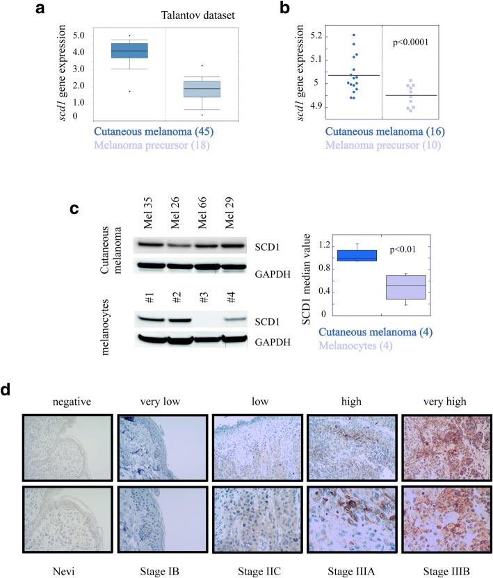
SCD1 is potentially useful for discriminating healthy tissue from melanoma. a) Geo Skin Cutaneous Melanoma Talantov dataset was analyzed for the expression of SCD1 by using Oncomine tool. Boxplot: cutaneous melanoma (n = 45); melanoma precursor (n = 18); b) mRNA expression of SCD1 was determined by qRT-PCR analyses in melanoma patients affected by different tumor stage. The samples are grouped for SCD1 gene by stage in melanoma precursor (n = 10) and cutaneous melanoma at different stage (n = 16); c) Western blotting analysis of SCD1 protein in four primary cell lines isolated from patients affected by cutaneous melanoma at different stage (upper panel) and four cell lines obtained from non tumoral tissue (bottom panel). Mel 26 stage IB; Mel 35 stage IIC; Mel 66 IIC and Mel 29 stage IIIC. On the right boxplots represent the quantification of SCD1 levels expressed as median value (fold-change = 1.9, p = 0.01); d) Representative images showing cellular variability for IHC staining of SCD1 protein in melanoma patients. Magnification 200X (upper) 400X (bottom)

