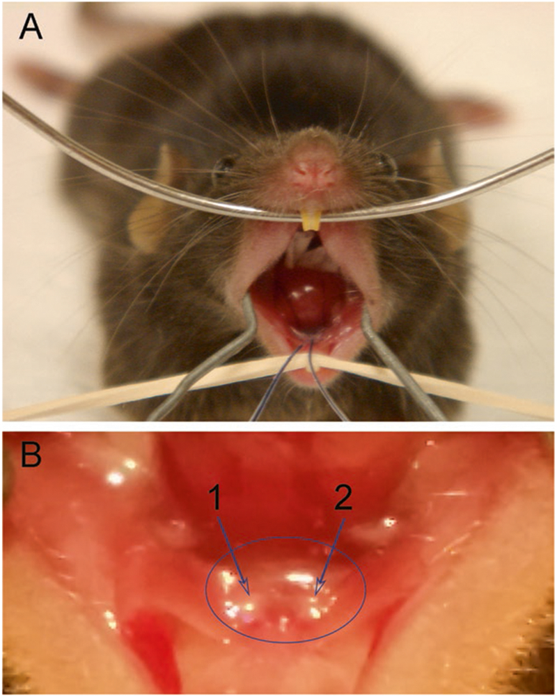
Fig. 4.

(a) Mouse with cannulas inserted into the orifices of Wharton’s duct (submandibular gland) prior to vector delivery (note needle and syringe attached to each cannula are out of the plane of this picture). Place the upper jaw of the mouse on a wire, i.e., teeth over the wire, and pull the lower jaw down with rubber band onto rack. Finally, expand cheeks with wire spring made from a bent paper clip. (b) Close-up view of right (“1 arrow”) and left (“2 arrow”) Wharton’s ducts in a mouse
