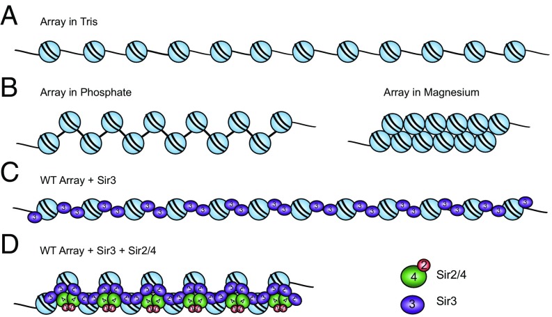
Fig. 5.
Model for a SIR chromatin fiber. (A) Diagram of a 12-mer array in low-salt Tris buffer. (B) Arrays in 20 mM phosphate buffer (pH 8.0; containing ∼40 mM Na+) are partially folded. Arrays in 1 mM MgCl2 buffer fold into 30-nm fibers. (C) Sir3 binds to arrays as a monomer, then subsequent dimerization via the Sir3 C-terminus bridges neighboring nucleosomes. (D) SIR proteins bind and condense WT arrays, though to a lesser extent than 30-nm fibers, with two molecules of Sir3 and likely one molecule of Sir2–4 per nucleosome.

