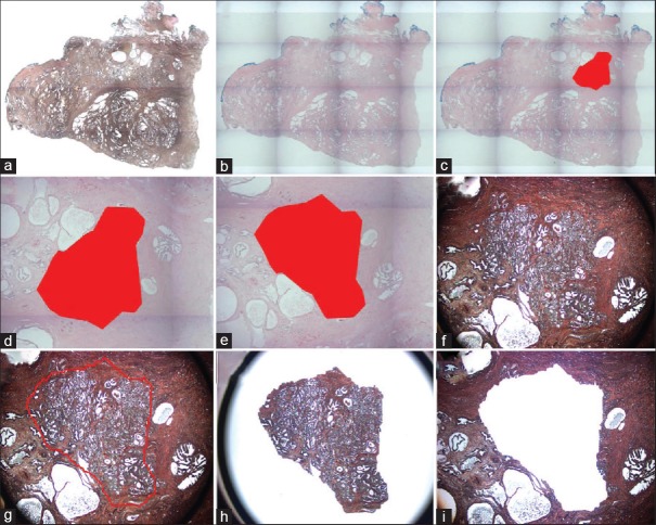
Figure 2.
Probabilistic pairwise markov model-laser capture microdissection (a) A low power image of an uncoverslipped Haemotoxylin and Eosin stained human prostate tissue section. (b) A low power image of the Haemotoxylin and Eosin stained tissue with the pseudo-coverslip. (c) Low power image of Probabilistic pairwise Markov model -identified tumor region annotated in red. (d) High power view of the Probabilistic pairwise Markov model area. (e) Pseudo-coverslipped image is rotated to fit. (f) Uncoverslipped sample at high power. (g) This image was imported into AutoScanXT and dissected by the ArcturusXT. (h) Laser capture microdissection cap showing area that was dissected. (i) The remaining tissue in the section is shown

