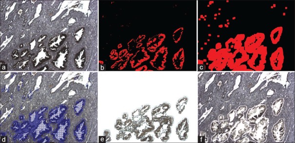
Figure 3.
ImageJ (a) Uncoverlipped immuno-stained human prostate tissue. (b) ImageJ binary mask for the DAB stained areas. (c) ImageJ dilation tool to increase coverage. (d) ImageJ imported into AutoScan software on the ArcturusXT. (e) Image of the laser capture microdissection cap after dissection. (f) Tissue that remains behind after laser capture microdissection

