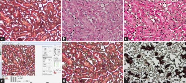
Figure 6.
eSeg with the Leica LMD7000 (a) Deparaffinized, Haemotoxylin and Eosin stained tissue on a PET membrane metal frame slide. (b) Same image as in (a), but with the xylenes pseudocoverslip. (c) eSeg applied to the image and the green contours around the target nuclei are shown. (d) The Leica display following transfer of pattern matching contours from eSeg integrated into the Leica LMD7000 workflow. (e) The Leica display console screen following transfer of pattern matching contours from eSeg. (f) The Leica dissection following eSeg-based computer-aided laser dissection on the LMD7000

