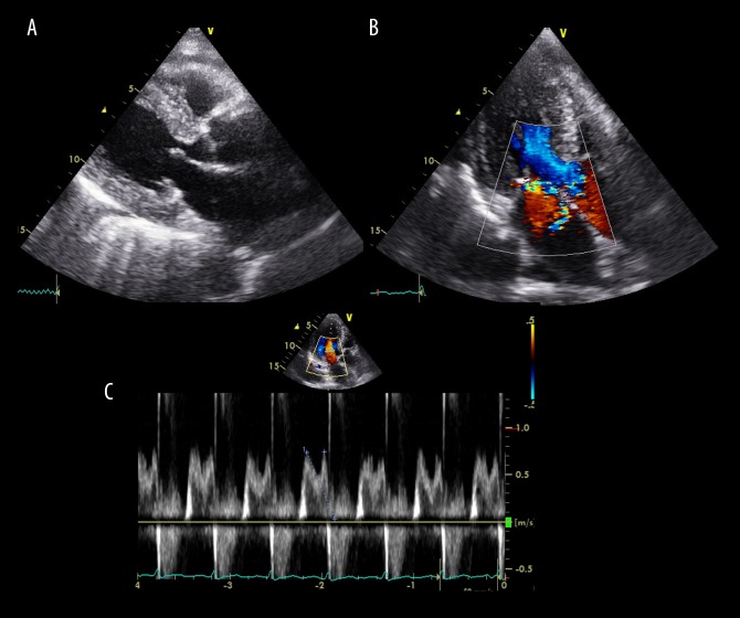
Figure 1.
Transthoracic echocardiography performed on admission. (A) Brightness mode image, (B) color Doppler image, and (C) pulse Doppler image. Ejection fraction of 70%, left atrial volume index of 66 mL/m2 and posterior wall thickening of 14 mm are shown. Mitral regurgitation (grades III to IV) and prolapse of posterior mitral leaflet P3 are revealed (B, C) without findings of vegetation and wall motion abnormalities (A).

