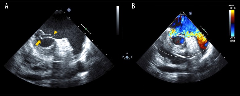
Figure 2.
First transesophageal echocardiography performed following transthoracic echocardiography. (A) Brightness mode image and (B) color Doppler image. Prolapse of posterior mitral leaflet P3 (arrow) and ruptured chordae tendineae (arrowhead) are shown without the finding of vegetation and hyperplasia of valves (A). The mitral regurgitation jet runs from the site of prolapse to left atrial appendage along the anterior leaflet (B).

