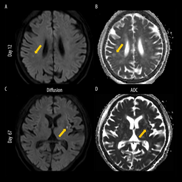
Figure 3.
Brain magnetic resonance imaging. (A) Diffusion-weighted image taken on day 12, (B) apparent diffusion coefficient (ADC) image taken on day 12, (C) diffusion-weighted image taken on day 67, and (D) ADC image taken on day 67. On day 12, the diffusion-weighted image shows high signal intensity in the right temporoparietal lobe (arrow, A) and ADC value of the lesion is low to equal (arrow, B). On day 67, the diffusion-weighted image shows high signal intensity in the left putamen (arrow, C) and ADC value of the lesion is low to equal (arrow, D). Images of these lesions are compatible with sub-acute to acute phase cerebral infarction, which is considered to be caused by cardiogenic emboli because of the involvements of both sides of the brain.

