Figure 3.

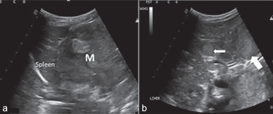
Two selected ultrasound images. (a) Large left adrenal mass (M). (b) Liver metastasis (small white arrow) and retroperitoneal lymph nodes (large white arrow).

Two selected ultrasound images. (a) Large left adrenal mass (M). (b) Liver metastasis (small white arrow) and retroperitoneal lymph nodes (large white arrow).