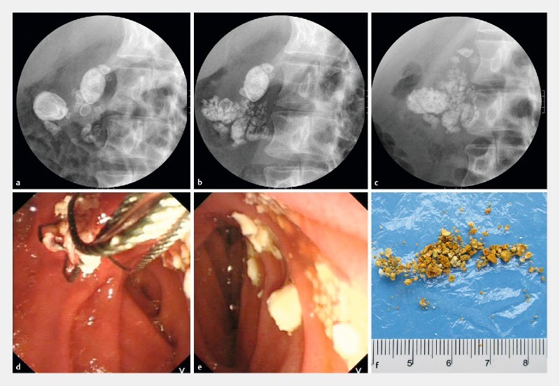
Fig. 1.

The process of pancreatic stone fragmentation and extraction. a A fluoroscopic image showing multiple stones in the head of pancreas before P-ESWL. The image was taken with the patient in a supine position tilting to right in an angle of 30°. b As shown in the X-ray. Parts of the stones were fragmented after the first P-ESWL. c After the second P-ESWL, the pulverized stones were less dense than they were prior to P-ESWL and spread all along the duct. The patient then received a third P-ESWL followed by ERCP. d During ERCP, plenty of white pulverized stones were extracted by extraction basket. e An endoscopic image showing extracted pulverized calculi. f Pancreatic stones identified from the stool of the patient.
