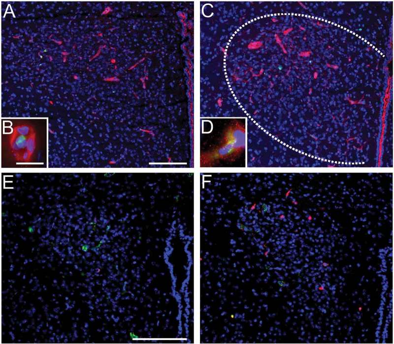
Figure 2.

LPS-induced systemic inflammation increase ICAM1-immunoreactivity and induces the recruitment of neutrophil granulocytes to the brain in the paraventricular nucleus (PVN). LPS (5 mg/kg i.p., 4.5h) induces ICAM1-immunoreactivity (red, a-d) in the PVN (c) compared to controls (a). Close association of ICAM1-immunoreactivity with myeloperoxidase (green) staining (a-d) is depicted in insets (b, d). In addition, LPS-stimulation (2.5 mg/kg 24h) increased the number of Ly-6B.2 alloantigen (clone 7/4) stained neutrophil granulocytes in the PVN (red, f) compared to controls (e). Von Willebrand factor (green, e-f) depicts brain vasculature. Blue DAPI staining visualizes the surrounding tissue (blue, a-f). Scale bars represent 100µm and 10µm in insets. The previously unpublished microphotographs were adapted from our own previous studies [15,53].
