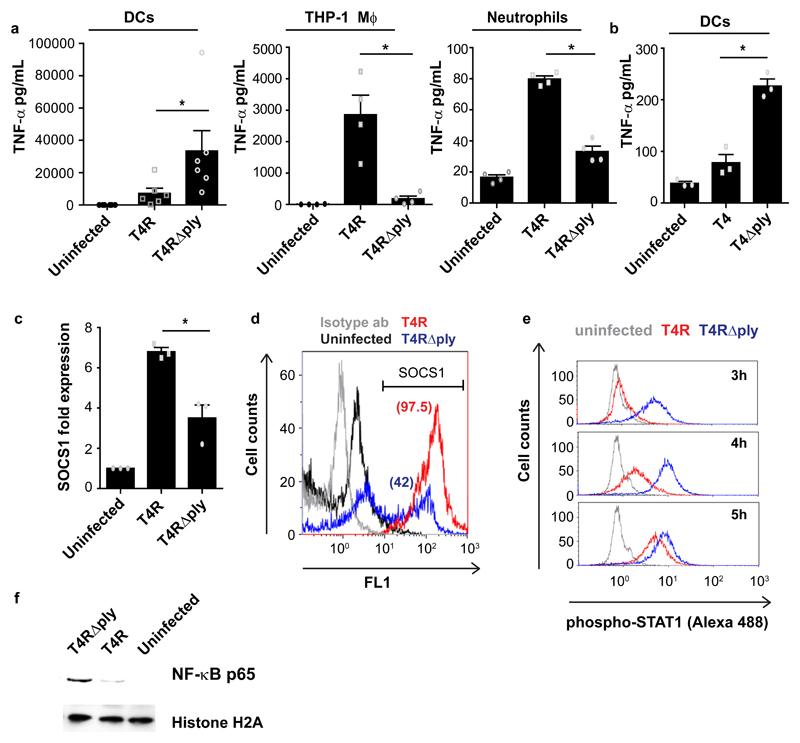
Fig. 1. Pneumolysin inhibits cytokine responses and inflammatory signalling in DCs by upregulating SOCS1.
(a) TNF-α secretion from human dendritic cells (DCs) (N=6), THP-1 macrophages (N=4), and primary neutrophils (N=4) upon infection with wild type strain T4R or its isogenic pneumolysin (PLY) mutant T4R∆ply. Data are mean±SEM. *P < 0.05 by Wilcoxon matched-pairs signed (two-tailed) rank test. (b) TNF-α secretion from DCs infected with encapsulated strains, T4 or T4∆ply (N=3 donors). Data are mean±S.E.M. P < 0.05 by Wilcoxon matched-pairs signed (two-tailed) rank test. (c) SOCS1 mRNA levels in T4R or T4R∆ply infected DCs at 9 hrs post infection (pi) (N=3 donors). Data are mean±S.E.M. P < 0.05 by paired two-tailed t test. (d) Flow cytometry histogram plot showing SOCS1 protein levels in T4R or in T4R∆ply infected DCs at 9 hrs pi. Percentage of SOCS1+ cells is indicated within the parenthesis. (e) STAT1 phosphorylation in T4R or in T4R∆ply infected DCs at 3-5 hrs pi. Data in d,e are representative of 3 independent experiments. (f) Western blot showing the levels of nuclear NF-κB (p65) in T4R or T4R∆ply infected DCs at 4 hrs pi. Histone H2A served as loading controls. Blots are representative of data from 2 independent experiments.

