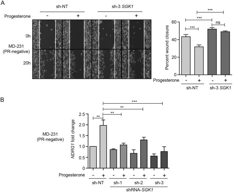
Figure 5.
Depletion of SGK1 renders breast cancer cells partially responsive to progesterone. A, cell migration assay upon depletion of SGK1 in MDA-MB-231 cells (PR-negative), in the presence and absence of progesterone treatment. Cells were monitored by a time-lapse wound-healing assay for 20 h. Cell migration from the 0- to 20-h time point is plotted as percentage wound closure, and the comparison was with respect to sh-NT clone. The bar plot indicates percentage wound closure for each of the clones, treated with or without progesterone, and the quantification is an average of three independent experiments performed in triplicates. B, transcript levels of NDRG1 have been analyzed in MDA-MB-231 cells (PR-negative) upon depletion of SGK1, in the presence and absence of progesterone stimulation. Data are plotted as -fold change of NDRG1 with respect to expression in untreated sh-NT cells and individual SGK1 knockdown clones. GAPDH was used as an internal normalization control. The analysis is representative of three independent experiments performed in triplicates. p value was calculated using Student's unpaired t test. **, p < 0.001; ***, p < 0.0001; ns, not significant. Error bars indicate S.D.

