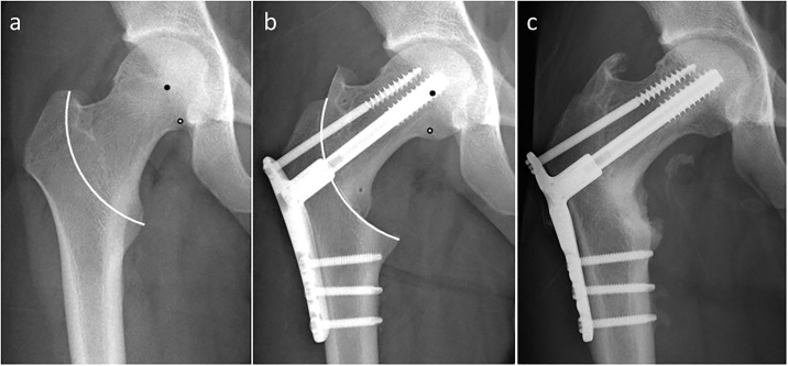
Fig 7. Radiographs of 20-year-old man who had steroid-associated osteonecrosis of the femoral head treated with curved intertrochanteric varus osteotomy.
a) Before and b) after the operation. White line indicates the osteotomy arc. Black points indicate the center of the femoral head and white points indicate the center of the osteotomy arc. The actual leg shortening was -2.0 mm, whereas the theoretical leg shortening was -1.6 mm.

