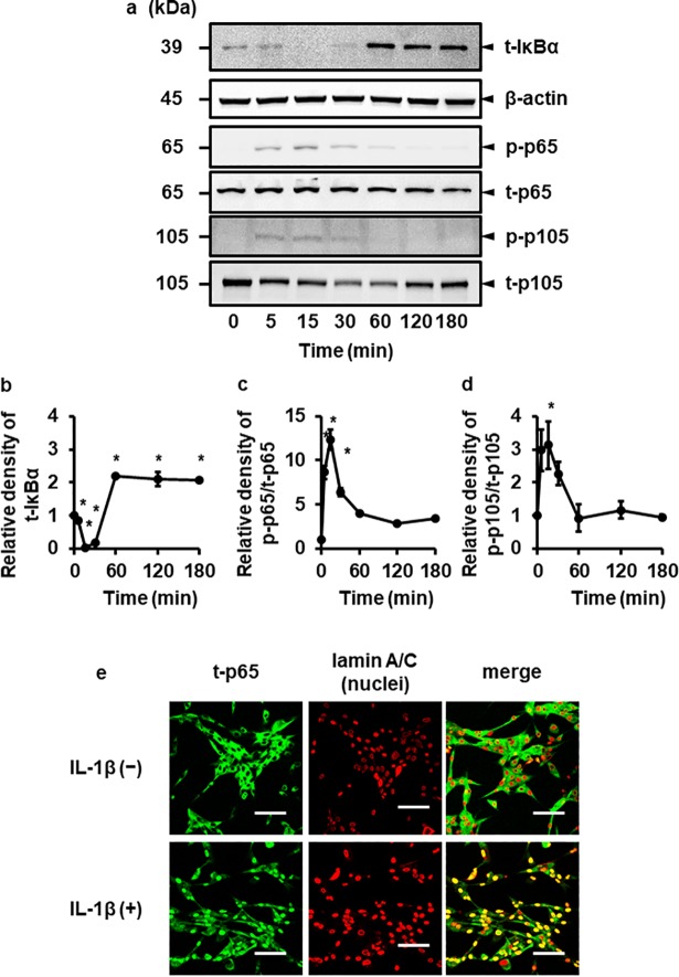
Fig 3. IL-1β induces the activation of canonical NF-κB pathway.
Canine melanoma cells were exposed to 100 pM IL-1β for the indicated time periods. At the end of the incubation, total (t-) IκBα, β-actin, total (t-) and phosphorylated (p-) forms of p65 and p105 were detected by immunoblotting. For the immunoblotting, cell lysate (10 mg protein) was used. Representative results of t-IκBα, β-actin, p-p65, t-p65, p105 and t-p105 expressions (a), and the relative density of t- IκBα (b), p-p65 (c) and p-p105 (d) compared to the results at time point 0 (lower panel) are depicted. Values are expressed as the mean ± S.E. of three independent experiments. *P < 0.05. (e) Canine melanoma cells were exposed to 100 pM IL-1β for 15 min. At the end of the incubation, t-p65 (green) and lamin A/C (red; nuclei) were detected by immunocytochemistry.

