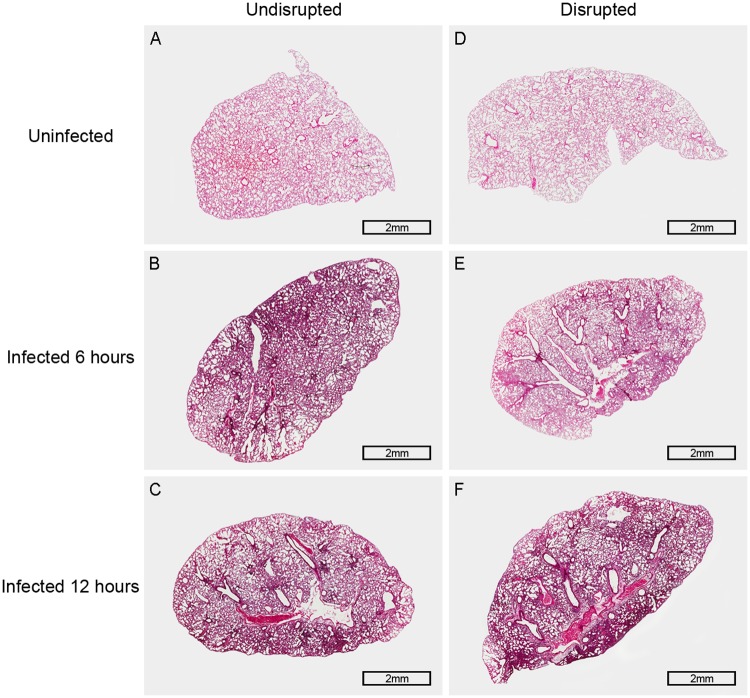
Fig 4. Histopathology of the lung in TLR4-deficient mice 6 hours and 12 hours after pneumococcal infection.
Representative pictures of H & E stained lung tissues: (A) The picture of the lung tissue in gut microbiota-undisrupted uninfected mice (n = 6 per group). (B and C) Pictures of the lung tissue in gut microbiota-undisrupted mice 6 hours and 12 hours after infection (n = 6 per group). (D) The picture of the lung tissue in gut microbiota-disrupted uninfected mice (n = 6 per group). (E and F) Pictures of the lung tissue in gut microbiota-disrupted mice 6 hours and 12 hours after infection (n = 6 per group). Original magnification, × 20; scale bar = 2 mm.

