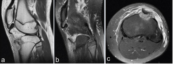
Figure 2.

Sagittal plane T1 (a), T2 (b), and axial T2 (c) cuts from the magnetic resonance imaging study demonstrate the tibial tubercle fracture as well as the distal patellar tendon rupture occurring off of its tubercle insertion. The tubercle fragment is rotated 180°, as seen in the axial plane (c).
