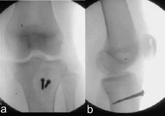
Figure 4.

Anteroposterior (a) and lateral (b) views of the left knee obtained from intraoperative fluoroscopy demonstrating fixation of the tibial tubercle fracture with the use of two fully-threaded cortical screws. After FiberWire fixation of the patellar tendon, patellar height appears restored on the lateral view as compared with the lateral view in Figure 1.a
