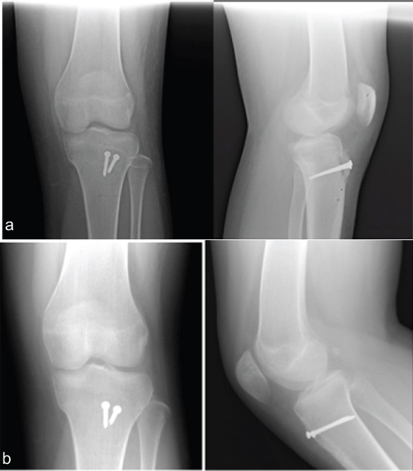
Figure 5.

Anteroposterior and lateral radiographs of the left knee at 4 weeks (a) and 5 months (b) postoperatively showing healing of the tibial tubercle fracture with the two cortical screws in proper position. On the lateral views, the position of the transverse drill holes can be visualized in the patella and the proximal tibia.
