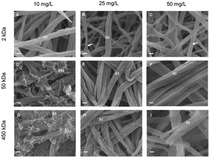
Figure 1:
SEM images of type I collagen fibrils mineralized for 7 days with 2k-10 (A), 2k-25 (B), 2k-50 (C), 50k-10 (D), 50k-25 (E), 50k-50 (F), 450k-10 (G), 450k-25 (H), and 450k-50 (I) PAA solutions. IM: fibrils with intrafibrillar mineralization. EM: extrafibrillar mineralization of fibrils. Arrows in (B) and (C): collagen fibrils not mineralized showing the characteristic banding pattern of native collagen. Scale bar: 200 nm.

