. 2018 Nov 26;7:e33718. doi: 10.7554/eLife.33718

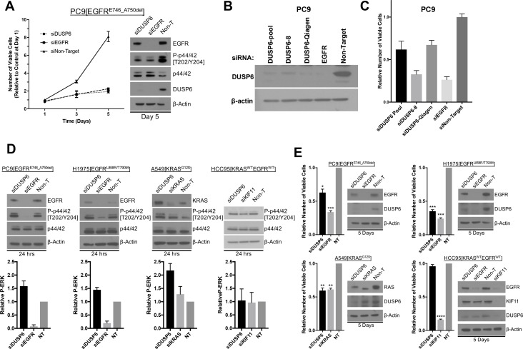
Figure 3. Knockdown of DUSP6 increases P-ERK and selectively inhibits LUAD cell lines with KRAS or EGFR mutations.

(A) Interference with DUSP6 RNA induces toxicity in PC9 cells. Pooled siRNAs for DUSP6, EGFR or a non-gene targeting control (Non-T) were transfected into PC9 cells (carrying an EGFR mutation) on day 0 and day 3, and the numbers of viable cells in each condition was measured with Alamar blue at the indicated time points and scaled to the Non-T condition at day 1 to measure the relative changes in numbers of viable cells. Experiments were done in biological triplicate with the average values presented ±SEM. Western blots were performed at the endpoint of the assay (day 5) to confirm reduced amounts of DUSP6 protein and measure levels of ERK and P-ERK (p42/44 and P-p42/44, respectively). (B–C) A siRNA that targeted the 5’ region of DUSP6 mRNA coding sequence (siDUSP6-Qiagen; different from siDUSP6-8 that targets the 3’ mRNA coding region), reduces levels of DUSP6 protein and decreases the numbers of viable cells. The indicated siRNAs (DUSP6-pool, DUSP6-8, DUSP6-Qiagen, EGFR and Non-Target) were delivered to PC9 cells, the levels of DUSP6 protein measured and the numbers of viable cells was determined as described for panel A. Experiments were done at least three times, and the average ±SEM is indicated for cell viability. (D) Interference with DUSP6 RNA acutely increases P-ERK levels. DUSP6 was knocked down in PC9 and H1975 cells (EGFR mutants), A549 cells (KRAS mutant), and HCC95 cells (KRAS and EGFR wild-type); levels of ERK and P-ERK were measured by Western blot 24 hr later. Relative P-ERK levels (ratio of phosphorylated to total levels normalized to actin) were determined by dosimetry and compared to the non-targeting control (NT) to quantify the relative increase after DUSP6 knockdown. Three independent western blots were performed and the average ±SEM is plotted. (E) Interference with DUSP6 RNA inhibits LUAD cell lines with activating mutations in genes encoding components of the EGFR/KRAS signaling pathway. Numbers of viable cells 5 days after knockdown of DUSP6 or knockdown of positive controls (EGFR, KRAS or KIF11) were assessed with Alamar blue and compared to the non-targeting controls to determine relative changes. Experiments were done in biological triplicate with the average values presented ±SEM. Western blots to monitor knockdown of target genes at Day 5 are also displayed. *p < 0.05, **p < 0.01, ***p < 0.001, ****p < 0.0001, NS = Not Significant.

Figure 3.

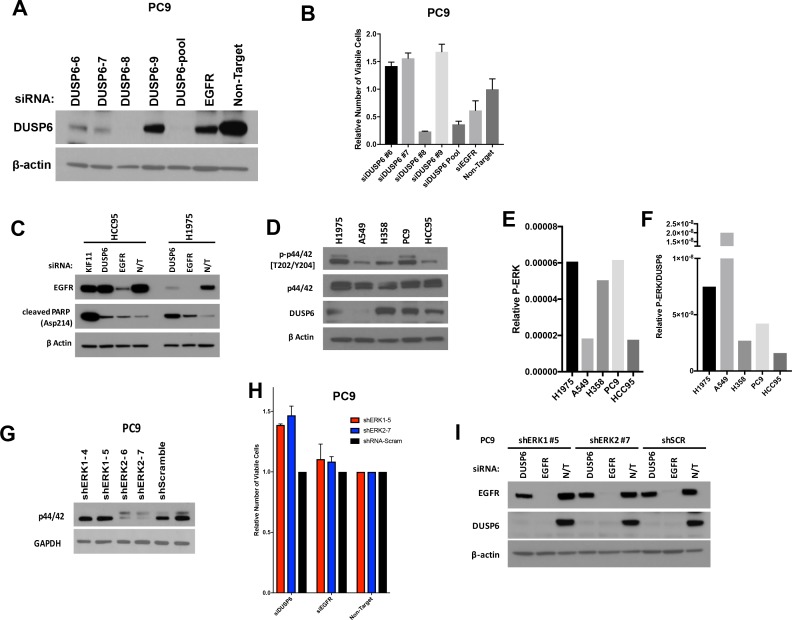
Figure 3—figure supplement 1. (A–B) Extensive knockdown of expression of DUSP6 with individual or pooled siRNAs is necessary to induce toxicity in PC9 cells.

Figure 3—figure supplement 1.

Individual siRNAs that comprise the pool of siRNA from Dharmacon (DUSP6-6,−7,–8 and −9) were transfected on days 0 and 3 into PC9 cells (carrying an EGFR mutation) at the same final concentrations as in Figure 3. Levels of DUSP6 protein were compared on day 5 in cells that received the individual DUSP6 siRNAs, DUSP6 siRNA pool, EGFR siRNA pool or a non-targeting control (Non-Target). (A) The number of viable cells was also measured with Alamar blue at day 5 and scaled relative to the sample that received the non-targeting siRNA to measure the relative change in viability. Experiments were done with at least three biological replicates; the average ±SEM indicated. (B) Only the DUSP6 siRNA pool and the siRNA DUSP6-8 reduced DUSP6 protein to nearly undetectable levels, with a concurrent decrease in viable cells. Conversely, less extensive knockdown of DUSP6, as seen after introduction of the other individual siRNAs (DUSP6-6,−7 and −9), was associated with an increase in viable cells. Experiments were done with biological triplicates; the average values presented ±SEM. (C) Interference with DUSP6 RNA specifically induces cleaved PARP in cells with RTK-RAS-ERK pathway mutations. Decreased numbers of viable cells were observed after knockdown of DUSP6 in cells with EGFR or KRAS mutations - but not those with wild-type versions of these genes - as in Figure 3C. As evidence that these effects are mediated in part by apoptosis, cleaved PARP is induced in EGFR mutant H1975 but not KRAS/EGFR wild-type HCC95 cells when DUSP6 is inhibited. KIF11 and EGFR siRNAs serve as positive controls for induction of apoptosis in HCC95 and H1975 cell lines, respectively. Western blots were performed at day 5 after transfections at day 0 and 3 as described above. (D–F) Comparison of basal levels of P-ERK and DUSP6. Western blots were performed with extracts of cell lines with (H1975, A549, H358 and PC9) and without (HCC95) EGFR or KRAS mutations. (D) Relative P-ERK levels (E) and P-ERK/DUSP6 levels (F) were determined and plotted for each line. Cell lines which display decreased numbers of viable cells after knockdown of DUSP6 have greater relative P-ERK and/or P-ERK/DUSP6 levels than those that do not show decreased numbers of viable cells. (G–I) ERK inhibition partially rescues PC9 cells from the toxic effects of DUSP6 knockdown. Lentiviral vectors containing shRNAs for either ERK1 or ERK2 were transduced into PC9 cells and puromycin treatment was used to establish stable cell lines as described in Figure 1. Resulting knockdown of ERK1 or ERK2 compared to scramble shRNA containing control cells was confirmed by western blot (G) PC9 cells with decreased ERK demonstrated increased relative numbers of viable cells after DUSP6 siRNA-mediated knockdown compared to control cells receiving scrambled siRNA, whereas no difference was observed after knockdown of EGFR. Experiments were done in at least biological triplicate with the average ±SEM indicated. (H) Knockdown of the intended target was confirmed by western blot (I) in stable cell lines. All experiments were performed as in Figure 3; measured with Alamar blue and western blots were performed at the end of the experiments (day 5).