. 2018 Nov 26;7:e33718. doi: 10.7554/eLife.33718

Figure 4. Treatment with the DUSP6 inhibitor BCI selectively kills LUAD cell lines with KRAS or EGFR mutation, implying a dependence on ERK-mediated signaling.

(A–B) BCI induces toxicity specifically in lung cancer cell lines with mutations in genes encoding components in the EGFR-KRAS-ERK pathway. (A) Eleven lung cancer cell lines were treated with increasing doses of BCI for 72 hr based on the reported effective activity of the drug (Shojaee et al., 2015). Cell lines could be assigned to three distinct groups: sensitive (red), intermediate (green) and insensitive (black). All sensitive cell lines contained either EGFR or KRAS mutations; the intermediate and insensitive cell lines were wild-type for genes encoding components of the EGFR-KRAS-ERK signaling pathway (as determined by the Sanger Cell Line Project and the Cancer Cell Line Encyclopedia [Barretina et al., 2012]). Experiments were done in biological duplicate with the average values presented ±SEM. (B) Crystal Violet stain of cells plated in the indicated doses of BCI or control (0 = 0.1% DMSO) for 72 hr. Sensitive cells with a KRAS mutation (H358 cells; denoted with red underlining) show a more pronounced decrease in cell number than do cells without oncogenic mutations in genes encoding components of the EGFR-KRAS-ERK pathway (H1648 cells; black underlining). Experiments were done in biological duplicate with a representative image shown. (C) BCI increases P-ERK levels specifically in BCI-sensitive cell lines. Sensitive lines (H358, PC9, H1975 and A549; red underlining) and insensitive lines (HCC95 and H1648; black underlining) were treated with the indicated doses of BCI or vehicle control (0.1% DMSO) for 30 min, and the levels of ERK (p44/p42) and P-ERK (P-p44/42 T202/Y204) assessed by Western blot. P-ERK appeared in the sensitive cells at low doses of BCI, but P-ERK levels did not increase in the insensitive cells at the tested doses of BCI. (D) Dosimetry plots from the experiment shown in panel. (C) (E–F) Cell lines sensitive to BCI are also dependent on P-ERK for survival. BCI-sensitive cells with oncogenic mutations in EGFR or KRAS (PC9 and H358, respectively; red underlining) and BCI-insensitive cells (H1648 and HCC95; black underlining) were treated with the indicated doses of the MEK inhibitor trametinib for 72 hr; viable cells were measured with Alamar blue and compared to cells receiving the vehicle control (0 = 0.1% DMSO). (E) Treatment with trametinib decreased P-ERK levels as determined by western blot. (F) The reduction in P-ERK corresponded to a greater decrease in viable cells in BCI-sensitive lines (red coloring), compared to BCI-insensitive cell lines (black coloring).

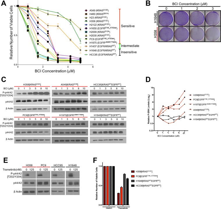
Figure 4.

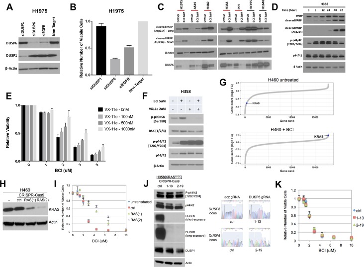
Figure 4—figure supplement 1. (A–B) Knockdown of DUSP6, but not DUSP1, decreases viability of LUAD cells.

Figure 4—figure supplement 1.

DUSP1 and DUSP6 siRNAs were introduced into H1975 cells as described in Figure 1C. On day 5, western blots were performed to confirm knockdown of appropriate proteins (A) and measured with Alamar blue was used to count viable cells, (B) relative to cells receiving non-targeting control siRNAs. Reduction of DUSP6, but not of DUSP1, decreases viable cells, suggesting DUSP6 is the primary mediator of BCI-induced toxicity. Experiments were done in at least biological triplicate, with the average ±SEM indicated. (C) BCI induces cleaved PARP specifically in lung cancer cell lines with mutations in genes encoding components in the EGFR-KRAS-ERK pathway. A subset of cell lines from Figures 4A5 categorized as sensitive (red line) and two insensitive (black line) - were treated with 3 uM BCI for 72 hr; induction of cleaved PARP was assessed by Western blot. Cleaved PARP is increased only in sensitive cell lines containing mutations in EGFR or KRAS. (D) Time course of cleaved PARP after BCI treatment in H358 cells in relation to pERK induction. 3 uM BCI increased P-ERK followed by cleavage of PARP, as determined by western blot at the indicated time points. (E–F) Decreased ERK activity partially rescues LUAD cells from BCI-induced toxicity. H358 cells received the indicated doses of BCI and the ERK inhibitor VX-11e for 72 hr; numbers of viable cells were determined by Alamar blue as in Figure 4A. Values for each line exposed to the VX-11e/BCI combination were normalized to results obtained only with VX-11e. Experiments were done in at least biological triplicate, with the average ±SEM indicated. Treatment of H358 cells with VX-11e decreased toxicity induced by BCI in a dose dependent manner, (E) corresponding to a decrease in downstream ERK activity as indicated by western blot for the ERK target RSK. (F) (G) Genome wide CRISPR-Cas9 screen in H460 cells reveals a dependence on KRAS for BCI sensitivity. The changes in abundance of guide RNAs are shown, revealing that a guide RNA targeting KRAS is depleted in control cells and enriched in the presence of BCI. (H) Validation of CRISPR-Cas9 screen. Two separate guide RNAs targeting KRAS (labeled 1 and 2) and a control gRNA targeting lacz (ctrl) were independently introduced into H460 cells, along with Cas9 in a lentiCRISPR v2 vector. Cell lines carrying these modifications and control cells were evaluated for KRAS depletion by western blot. The same cell lines were evaluated for their sensitivity to BCI in a dose response curve (I). Viable cell numbers are plotted relative to each line in the absence of BCI (set to 1.0) (J) H358 cells deficient in DUSP6 are responsive to BCI. Clones derived from H358 cells carrying a control (ctrl) guide RNA or two independent DUSP6 guides (1-13, 2-19) were evaluated by western blot for abundance of the indicated protein (left) and for successful targeting of the DUSP6 locus by DNA sequencing (right). DUSP6 protein is absent in the two clones. (J) H358 cells deficient for DUSP6 and cells targeted with a control gRNA were evaluated for sensitivity to BCI in a dose response curve. (K) Viable cell numbers are plotted relative to each independent line in the absence of BCI (set to 1.0). Results are representative of 3 independent experiments.