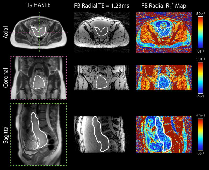
Figure 3.

Representative in vivo placenta images and R2* maps of a subject with normal pregnancy at 16+2 weeks gestational age acquired using free-breathing (FB) radial MRI at 3 T. Axial, coronal and sagittal views are shown. The placenta is delineated by a white contour.
