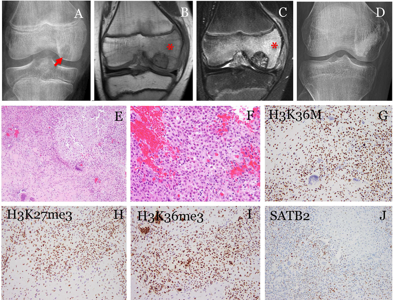
Fig. 1.
A representative case of chondroblastoma with positive H3K36M immunostaining. The patient was a 11-year-old male. (A) Radiograph showed an epiphyseal lytic tumor(arrow). (B) MRI T1- and (C) T2-weighted fat-suppressed images showed the tumor surrounded by edema (*). The tumor was curetted and cemented. The patient was followed for 58 months and (D) radiograph showed no recurrence. (E) Histologically, the tumor was composed of sheets of uniform cells with focal chondroid differentiation and scattered multi-nucleated giant cells (200x). (F) The tumor cells were oval to polygonal with well-defined pink cytoplasm and cleaved nuclei (400x). (G) The tumor cells showed strong nuclear staining of H3K36M (200x). Immunostaining for methylated histones, (H) H3K27me3 and (I) H3K36me3, showed heterogeneous staining in both the mononuclear tumor cells and the multi-nucleated giant cells (200x). (J) SATB2 was positive in a subset of the tumor cells (200x).

