Fig. 3.
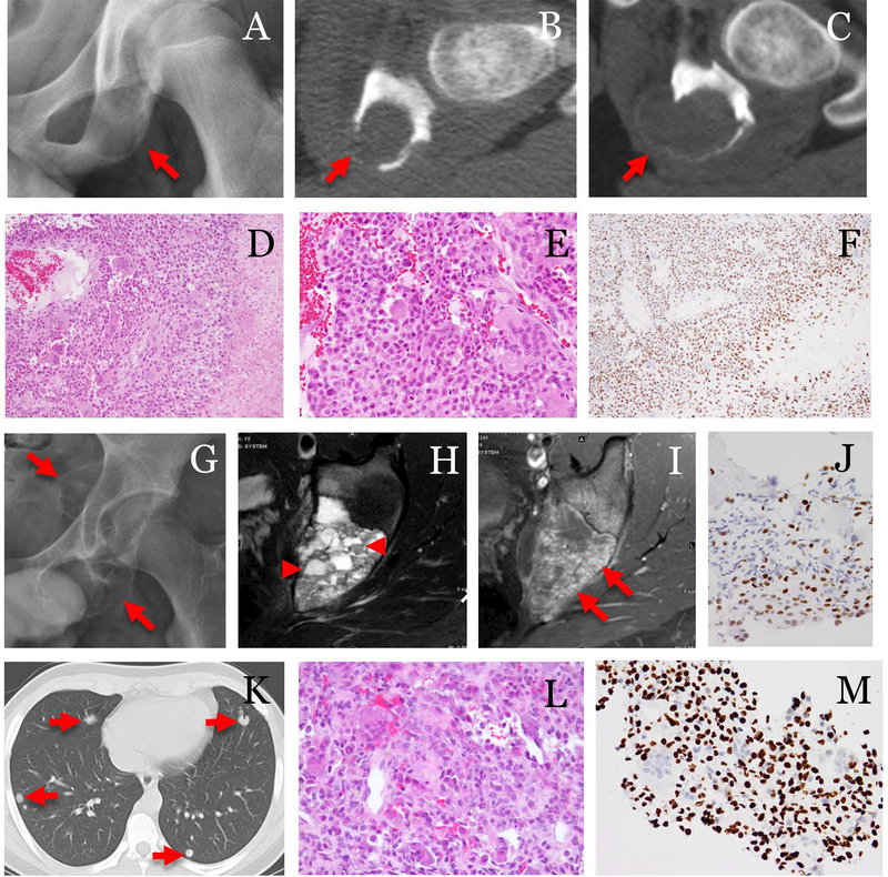
A case of chondroblastoma with lung metastasis. The patient was a 13-year-old male. (A) Pre-op radiograph showed a lytic ischial lesion(arrow). (B, C) Serial CT exams in 2 months showed increased size and cortical destruction of the tumor(arrows). The tumor was treated by embolization, curettage and bone grafting. (D) Histologically, the tumor was composed of sheets of uniform cells with chondroid differentiation, aneurysmal bone cyst-like change, and frequent multi-nucleated giant cells (100x). (E) The tumor cells showed typical morphology of chondroblasts (400x) and (F) were strongly positive for H3K36M (200x). Three months after curettage, (G) radiograph showed recurrent tumor (arrows). (H) MRI T2-weighted, and (I) T1-weighted FS post-contrast images showed fluid levels (arrowheads) and enhancing components(arrow) in the tumor. (J) The recurrence was biopsied and tumor was again positive for H3K36M immunostaining. At 11 months after the surgery, (K) CT showed pulmonary nodules(arrows). (L) Needle core biopsy of a lung nodule confirmed metastatic chondroblastoma, which showed similar morphology as that seen in the primary tumor (400x) and (M) tumor cells were positive for H3K36M immunostaining (400x).

