
Figure 1. Confirmation of the likely pathogenic VUSMYL3(170C>A) uncovered in an asymptomatic carrier.
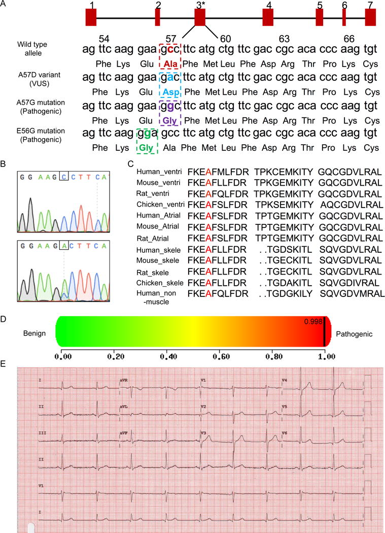
(A) Schematic illustration of the nucleotide and amino acid position of the VUS (NM_000258.2:c.170C>A, NP_000249.1:p.Ala57Asp) and two known pathogenic mutations (A57G, E56G) in the MYL3 gene. The red rectangles represent exons 1-7. Asterisks displays VUSMYL3(170C>A) position. Numbers 54-66 annotate amino acids positions. (B) Sanger sequencing confirmation of the VUSMYL3(170C>A). Upper panel: Sequencing result of a healthy control individual, lower panel: sequencing result of the heterozygous VUSMYL3(170C>A) carrier. Nucleotide position of the VUSMYL3(170C>A) is displayed by a framing square. (C) Alignment of regions flanking the VUSMYL3(170C>A) (red font) in MYL3 protein showing evolutionary conservation of the mutated residue across species and isoforms. (D) A prediction score of the VUSMYL3(170C>A) predicted by the in silico tool PolyPhen-2. (E) The VUSMYL3(170C>A) carrier’s ECG recording.