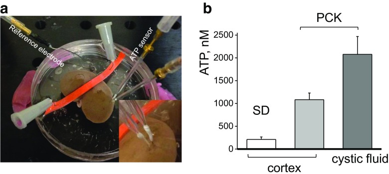
Fig. 1.
ATP levels in PCK cystic fluid and cortical tissues of the isolated kidneys from PCK and Sprague-Dawley (SD) rats. a Enzymatic biosensor setup for detection of ATP in the isolated kidney tissues. ATP and Null (to subtract noise and non-selective interferences) biosensors were inserted into the kidney cortical layer and connected to a dual channel potentiostat for real-time amperometry recordings. Inset shows a close-up image with sensors’ tips inserted into a cortex of the freshly isolated kidney from PCK rat. b ATP concentrations were detected using enzymatic biosensors in the cortical tissues of the freshly extracted kidneys from PCK and SD rats and isolated cystic fluid from PCK rats (N = 4 rats for each group)

