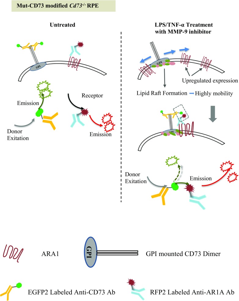
Fig. 8.
Illustration of FRET for detecting CD73 ARA1 association. Anti-CD73 antibody, labeled with EGFP2, was used as the donor fluorescent molecule (excitation maximum/emission maximum, 483/506 nM). Anti-ARA1 antibody, labeled with RFP, was used as the receptor fluorescent molecule (555/584 nM). When excited at 483 nM, EGFP2 generates green fluorescence with an emission wavelength spectrum ranging from 470 to 600 nm, which overlaps RFP’s excitation wavelength spectrum ranging from 470 to 610 nM. In untreated RPE cells, ARA1 is poorly expressed, and CD73 is inert without being localized to lipid rafts. These proteins are separated in the cell membrane. When the donor fluorescence excitation wavelength (483 nM) was applied, the emission energy from the donor could not be utilized by the receptor due to relatively large distance, and red fluorescence could not be visualized. Following LPS/TNF-α treatment, ARA1 expression was significantly increased, and the lipid rafts provided CD73 with high mobility. These facilitated the association of CD73 with ARA1. Once closely interacted, donor-emitted energy can be transferred to the receptor, and excited red fluorescence emission. It could be determined whether CD73 and ARA1 were closely interacted by detecting FRET’s fluorescence intensity

