Fig. 9.

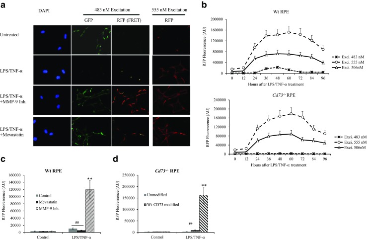
FRET assay for determining associated CD73-ARA1. In vitro cultured wild-type RPE and Cd73−/− RPE modified with Wt- and Mut-CD73 were differentially treated and subjected to FRET assay. Briefly, EGFP2-labeled anti-CD73 and RFP-labeled anti-ARA1 antibodies were added to RPE cells; the fluorescence from FRET receptor of GFP was detected by fluorescence microscopy (a) and a fluorometer (b–d). a Wt-RPE cells were treated with LPS/TNF-α and other factors. Green and red fluorescence were investigated by fluorescence microscopy. b The fluorescence intensity of RFP, excited by varied wavelengths at different times after LPS/TNF-α treatment, was evaluated by a fluorometer (n = 6) in Wt- (upper panel) and Cd73−/− RPE (lower panel) cells. c FRET-induced fluorescence of RFP in Wt-RPE at 48 h after treatment (n = 6). d FRET-induced fluorescence of RFP in Cd73−/− RPE modified with Wt- and Mut-CD73 at 48 h after treatment (n = 6)
