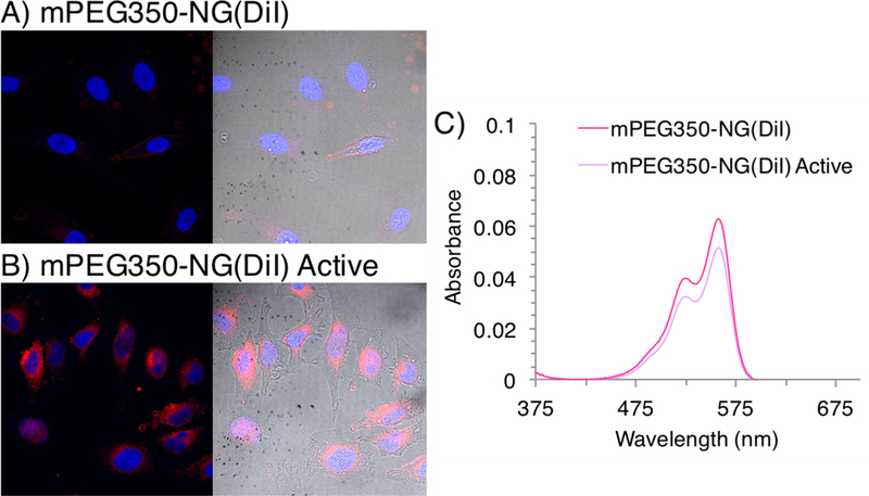
Figure 7.

Confocal microscopy images using a 40× objective of composite (left) with nucleus (blue 405 nm) and DiI-loaded NG (red, 540 nm), and composite with brightfield (right) image overlays of HeLa cells after 2-hour incubation with A) mPEG350-NG(DiI) and B) mPEG350-NG(DiI) Active. C) UV-visible absorbance spectra of DiI encapsulation before and after MMP-9 activation. Dye release calculated from decrease in absorbance at λmax 558 nm.
