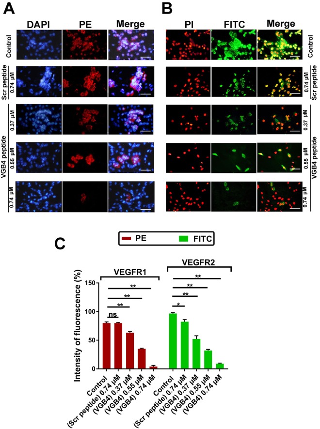
Figure 3.
The binding properties of VGB4 to VEGFR1 and VEGFR2 using immunofluorescence. HUVECs were treated for overnight with VGB4, fixed and stained with (A) anti-VEGFR1 –primary antibody which was detected with PE-labeled goat anti-mouse IgG secondary antibody, and (B) anti-VEGFR2 primary antibody which was detected with FITC-labeled rabbit anti-mouse IgG secondary antibody. (C) Graph representing the percent of fluorescence intensity in treated groups compared to control and scr peptide. All data were represented as mean ± SD of three experiments. *P ≤ 0.05, **P ≤ 0.001 versus control. All images were taken by Olympus fluorescence microscope and magnification 400×; bar = 50 µm.

