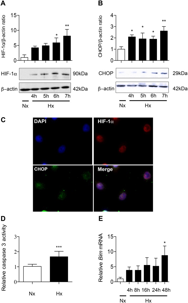
Figure 4.
Hypoxia induces HIF-1α, CHOP and apoptosis in alveolar epithelial cells exposed to hypoxia. Primary rat AECs were exposed to normoxia (Nx) (21% of O2) or hypoxia (Hx) (1.5% of O2) for increasing times (4–24 h). Protein levels of HIF-1α (A) and CHOP (B) were evaluated by western blotting and were normalized to the corresponding β-actin signal. Rat AECs were exposed to 6 h-hypoxia and immunolabeled for HIF-1α (red) and CHOP (green). DAPI was used to stained nucleus (blue) (C). The activity of effector caspase 3 was evaluated by enzymatic assay (D). Expression of the pro-apoptotic marker Bim was evaluated by RT-qPCR (E). n = at least 5 independent AECs cultures. Data were submitted to a Kruskal-Wallis one-way analysis of variance followed by a Dunn’s multiple comparison tests. *P < 0.05, **P < 0.01 and ***P < 0.001 represent a significant difference as compared with normoxic condition.

