
Figure 7.
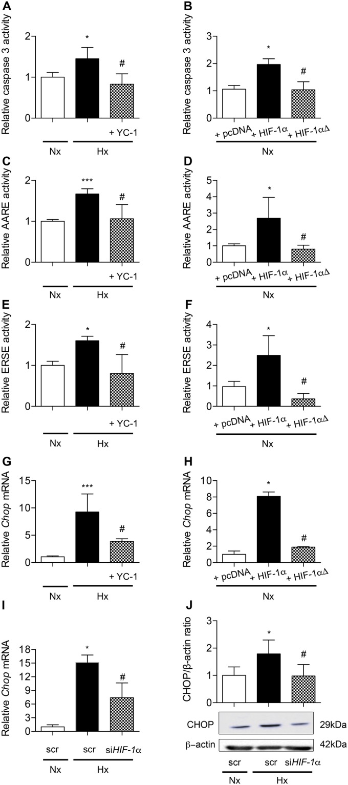
HIF-1α is involved in ER-stress induced CHOP-dependent apoptosis in alveolar epithelial cells. Caspase 3 activity was evaluated in primary rat AECs treated or not with the HIF-1α inhibitor YC-1 (10 μM) and exposed to normoxia (Nx) (21% of O2) or hypoxia (Hx) (1.5% of O2) for 24 h (A). A549 cells were transfected with either an empty pcDNA3.1 vector or a plasmid encoding HIF-1α or a mutated HIF-1α (HIF-1αΔ) unable to transactivate. Caspase 3 activity was measured 48 h after transfection (B). ATF4 (C) or ATF6α/XBP1s (E) relative transcriptional activities were evaluated in primary rat AECs treated or not with YC-1 and exposed to normoxia or hypoxia for 6 h. In A459 cells co-transfected with either an empty pcDNA3.1 vector or a plasmid encoding HIF-1α or a mutated HIF-1αΔ, ATF4 (D) or ATF6α/XBP1s (F) relative transcriptional activities were measured 48 h after transfection. CHOP mRNA expression was evaluated by RT-qPCR in primary rat AECs treated or not with YC-1 and exposed to normoxia or hypoxia for 6 h (G). CHOP mRNA expression was evaluated by RT-qPCR in A459 cells transfected with either an empty pcDNA3.1 vector or a plasmid encoding HIF-1α or mutated HIF-1αΔ 48 h post-transfection (H). A549 cells were transfected with HIF-1α siRNA or scrambled (scr) siRNA, and exposed to hypoxia (0.5% of O2) for 24 h. CHOP expression was evaluated by RT-qPCR (I) and western blotting (J). n = at least 5 experiments. Data were submitted to a Kruskal-Wallis one-way analysis of variance followed by a Dunn’s multiple comparison tests. *P < 0.05, ***P < 0.001: significantly different from normoxic control value (A,C,E,G), from value in normoxic scrambled-transfected cells (I–J) or from value in normoxic pcDNA3.1-transfected cells (B,D,F,H). #P < 0.05, significantly different from value in untreated hypoxic cells (A,C,E,G), in hypoxic cells transfected with scrambled siRNA (I–J), or in hypoxic cells transfected with pcDNA3.1 (B,D,F,H).