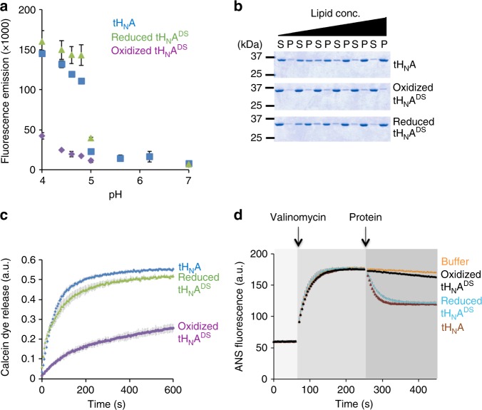
Fig. 3.
Modulating the conformation of the BoNT-switch using an engineered disulfide bond trapping. tHNADS was analyzed by four independent assays: a ANS binding; b liposome association: liposome co-sedimentation was conducted by incubating the WT tHNA or tHNADS with different concentrations of liposomes containing 20% DOPS and 80% OBPC. Supernatant (S) and pellet (P) fractions were analyzed by SDS-PAGE. Quantification of protein band intensities are shown in Supplementary Fig. 12. Uncropped images of gels are shown in Supplementary Fig. 13; c calcein dye release; and d membrane depolarization assay. 5 mM TCEP was added for the reaction of reduced tHNADS and no reducing agent was used for wild type or oxidized tHNADS. The data are presented as mean ± S.D., n = 3

