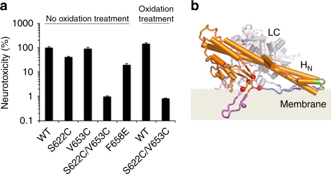
Fig. 5.

The BoNT-switch modulates neurotoxicity of BoNT/A1. a Neurotoxicity of BoNT/A1 variants was analyzed using the MPN assay. The paralytic half-times were determined and converted to the corresponding concentrations of wild-type BoNT/A1 using a concentration–response curve. The resulting toxicities were expressed relative to wild-type BoNT/A1 (n = 3–6, ± S.D.). b Proposed model for insertion of BoNT/A1 into lipid bilayer. The β2/β3 loop and the two putative membrane-interacting segments, F667–I685 and R827–R836, are colored in magenta, blue, and green, respectively. The inter-chain disulfide bond and four conserved carboxylates (D625, D629, E666, E670) are drawn as salmon and red spheres, respectively
